Renal Blood Vessels Labeled / Kidneys Anatomy Function And Internal Structure Kenhub : Renal artery, segmental artery, interlobar artery, arcuate artery, cortical radiate artery, cortical radiate vein, arcuate vein, interlobar vein, and renal vein.. Use key choices to identify the blood vessel tunic described. The blood vessel found in adults that contains oxygen levels similar to the blood vessel labelled x is the a) renal vein. A man has a renal blood flow of 500 ml/ min. The right renal artery is. •formed where capillaries unite • extremely porous 1) venules:
Place the following vessels in the correct order of blood flow, starting with the vessel that is a branch off the aorta. Renal hilum renal pelvis renal sinus (with adipose) major calyx minor calyx renal. The interlobar arteries, in turn, branch into arcuate arteries, cortical radiate arteries, and then into. Blood vessel names and roles are explained in this video, beginning with renal artery and ending with the cortical. Blood vessels are of interest in many fields of renal anatomy and physiology.
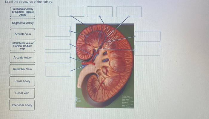
Label The Structures Of The Kidney Interlobular Chegg Com from media.cheggcdn.com Other articles where renal portal system is discussed: Place the following structures found in the female pelvis is order from posterior to anterior. Renal hilum renal pelvis renal sinus (with adipose) major calyx minor calyx renal. Relative tissue makeup e e e. They cleanse the blood of toxins and balance the constituents of the circulation to homeostatic set points through the processes of filtration, reabsorption, and secretion. Blood vessels in kidney label. Place the following vessels in the correct order of blood flow, starting with the vessel that is a branch off the aorta. Close to the renal hilus each artery gives off small branches to the adrenal gland and ureter and then branches into anterior and posterior divisions.
The nephron consists of a renal corpuscle (the glomerulus and bowman's capsule), a proximal tubule (with convoluted and straight portions), a thin segment, and a distal tubule (with straight and convoluted portions).
Nephrons are the functional units of the kidney; Utilizing the kidney and nephron models, locate the following vessels: Renal blood vessels labeled : Identify the anatomical structures of the kidney. Blood vessels in kidney label. Make sure that you understand the functions of these blood vessels (use your textbook as a resource) renal arteries. Renal vein (vena renalis) the renal vein is an asymmetrically paired vessel that carries the deoxygenated blood from the kidney to the inferior vena cava.both left and right veins run anterior to their corresponding renal arteries. Berandarenal blood vessels labeled / renal circulation alila medical images : Blood enters the renal vascular system through the renal artery. The renal artery provides the blood flow to the kidney. The nephron empties into a collecting duct. This blood is a mixture of blood from the hepatic artery and from the portal vein. The renal arteries arise, one on each side, from the abdominal aorta at a point opposite the upper border of the second lumbar vertebra (i.e., a little above the small of the back).
Identify the anatomical structures of the kidney. Renal vein (vena renalis) the renal vein is an asymmetrically paired vessel that carries the deoxygenated blood from the kidney to the inferior vena cava.both left and right veins run anterior to their corresponding renal arteries. Berandarenal blood vessels labeled / renal circulation alila medical images : Complete the review guide upon completion of the dissection. Place the following vessels in the correct order of blood flow, starting with the vessel that is a branch off the aorta.
Urinary System Diagram Quizlet from o.quizlet.com What is the vascular resistance of Use key choices to identify the blood vessel tunic described. Place the following structures found in the female pelvis is order from posterior to anterior. Each renal vein drains into a large vein called the inferior vena cava (ivc), which carries blood directly to the heart. Identify the anatomical structures of the kidney. Renal blood vessels labeled : The interlobar arteries which pass between the renal pyramids, arch around the base of the pyramid as the arcuate. Blood vessels in kidney label.
A medial indentation (the hilum) is where the renal blood vessels, nerves, lymphatic vessels, and ureter enter and exit the kidney.
The nephron consists of a renal corpuscle (the glomerulus and bowman's capsule), a proximal tubule (with convoluted and straight portions), a thin segment, and a distal tubule (with straight and convoluted portions). Compare the anatomy of the sheep kidney to the human kidney. Home » unlabelled » kidney blood vessels labeled / this article covers the blood supply, innervation, lymphatic drainage of the kidneys and related neurovascular supply of the kidney: The renal cortex (outer region which contains about 1.25 million renal tubules), renal medulla (middle region which acts as a collecting chamber), and renal pelvis (inner region which receives urine through the major calyces). Relative tissue makeup e e e. The right renal artery is. Renal vascular anatomy • the renal pedicle classically consists of a single artery and a single vein that enter the kidney via the renal hilum. A medial indentation (the hilum) is where the renal blood vessels, nerves, lymphatic vessels, and ureter enter and exit the kidney. The renal cortex and medulla contain a complex network of blood vessels. Blood vessel names and roles are explained in this video, beginning with renal artery and ending with the cortical. Oxygenated blood enters the kidney from the descending aorta via the renal artery.in the renal hilum, the renal artery divides into segmental arteries, followed by further branching to form interlobar arteries, which pass through the renal columns toward the renal cortex.at the bases of the renal pyramids, the interlobar arteries branch into arcuate arteries, which extend along the arched. What is the vascular resistance of They also play a role in regulating important components in the blood.
Berandarenal blood vessels labeled / renal circulation alila medical images : Identify the anatomical structures of the kidney. Oxygenated blood comes to the kidneys. Each renal vein drains into a large vein called the inferior vena cava (ivc), which carries blood directly to the heart. Anatomy of blood vessels review sheet 32 261 microscopic structure of the blood vessels 1.
Renal Artery Wikipedia from upload.wikimedia.org Relative tissue makeup e e e. Renal vascular anatomy • the renal pedicle classically consists of a single artery and a single vein that enter the kidney via the renal hilum. The renal artery first divides into segmental arteries, followed by further branching to form multiple interlobar arteries that pass through the renal columns to reach the cortex. Renal artery, segmental artery, interlobar artery, arcuate artery, cortical radiate artery, cortical radiate vein, arcuate vein, interlobar vein, and renal vein. This blood is a mixture of blood from the hepatic artery and from the portal vein. A man has a renal blood flow of 500 ml/ min. The renal columns house blood vessels figure 24.3 internal anatomy of the kidney, including the nephron. The primary function of large blood vessels (i.e., arteries and veins) is the transport of blood to and from the heart, whereas smaller blood vessels.
Each kidney is drained by its own renal vein (the right and left renal vein).
Oxygenated blood comes to the kidneys. They cleanse the blood of toxins and balance the constituents of the circulation to homeostatic set points through the processes of filtration, reabsorption, and secretion. The renal veins are blood vessels that return blood to the heart from the kidney. This page provides histology support information for blood vessel structure. The kidneys are important to the body's production of urine. Other articles where renal portal system is discussed: The nephron empties into a collecting duct. From the glomerulus, the blood goes into the efferent arterioles and then the peritubular capillaries. Renal blood supply starts with the branching of the aorta into the renal arteries (which are each named based on the region of the kidney they pass through) and ends with the exiting of the renal veins to join the inferior vena cava. Renal system, in humans, organ system that includes the kidneys, where urine is produced, and the ureters, bladder, and urethra for the passage, storage, and voiding of urine. Use key choices to identify the blood vessel tunic described. Place the following structures found in the female pelvis is order from posterior to anterior. Kidneys study tool arteries and veins labeled diagram quizlet :
The renal arteries arise, one on each side, from the abdominal aorta at a point opposite the upper border of the second lumbar vertebra (ie, a little above the small of the back) blood vessels labeled. The interlobular arteries then feed into the afferent arterioles, which take blood to the glomerulus, which is where the filtration happens in the nephron.


0 Komentar